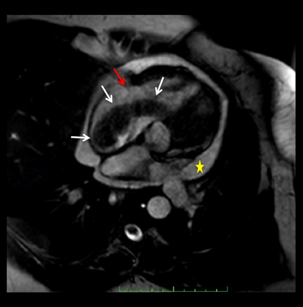
Figure 7.

Static frame of a cine steady state free-precession (SSFP) 4 chamber cardiac MR image. Low signal intensity, tubular shaped, worm-like mass (white arrow) is visualized extending from the right atrium through the tricuspid valve (red arrow) to the right ventricle. Pericardial effusion (yellow star). The mass does not invade the myocardium.
