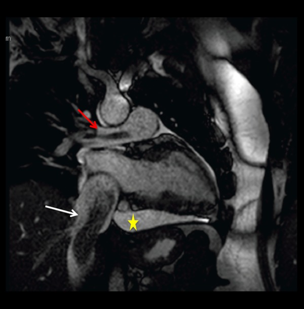
Figure 9.

Static frame of a cine SSFP oblique vertical long axis cardiac MR image. Low signal intensity, tubular shaped, worm-like mass (white arrow) is seen within the IVC. A smaller tubular shaped mass of similar characteristics is visualized within the right pulmonary artery (red arrow), related to extension of tumor to the pulmonary vasculature. Pericardial effusion (yellow star).
