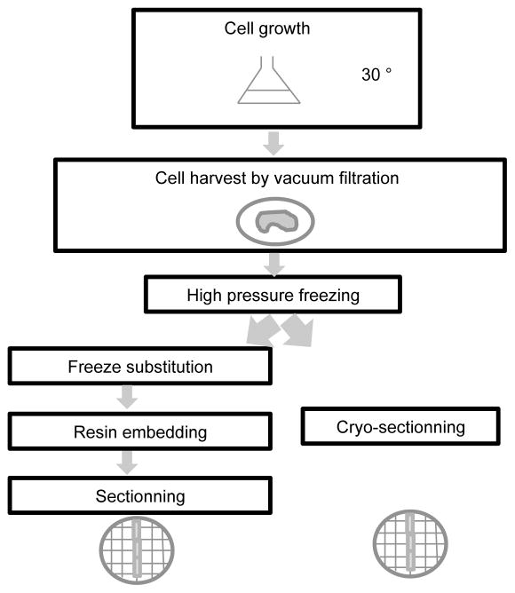
Figure 1. Schematic of the procedure used for sample preparation for electron microscopy analysis.
After cell growth, the cells are harvested by vacuum filtration and vitrified by high pressure freezing. Two strategies can then be employed. Either the vitrified cells are cryo-sectioned right before observation in the scope (or storage) or the cells are freeze substituted, embedded in resin at room temperature and sectioned.

