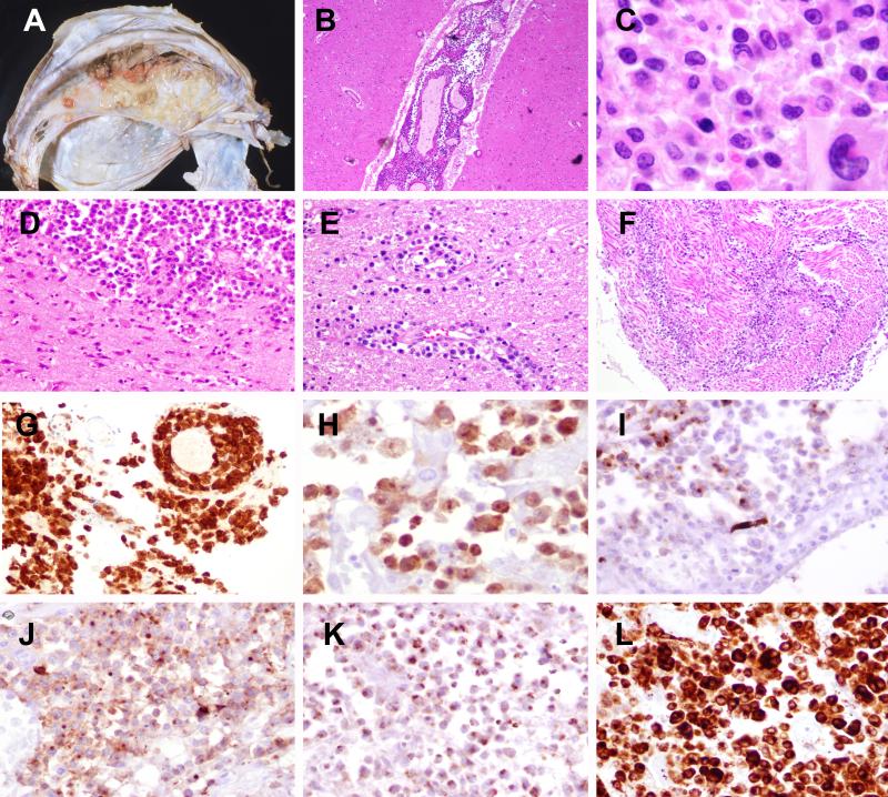
Figure 1. Anaplastic large cell lymphoma, ALK-positive (Case 16).
A) Multiple tan-white lesions are attached to the left side of falx dura. B) The leptomeninges are extensively involved. C) The cells are medium to large with irregular nuclei; occasional larger cells have eccentric kidney or horse shoe shaped nuclei and abundant cytoplasm consistent with “hallmark cells”. D) Parenchymal involvement was also seen along with perivascular infiltrates (E) as well as extensive spinal and nerve root involvement (F). The cells are positive for CD30 (G), ALK, nuclear and cytoplasmic (H), focal EMA (I), CD43 (J), TIA (K) and granzyme-B (L).

