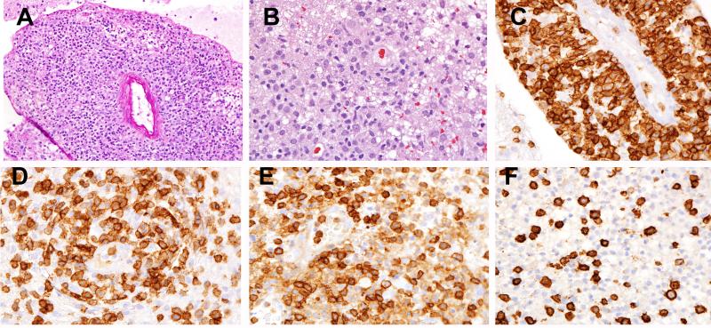
Figure 4. PTCL, NOS (Case 8).
(A) Marked perivascular infiltration is present. (B) The infiltrate is composed of small to medium atypical lymphocytes with significant background gliosis and abundant histiocytes. The atypical cells are strongly positive for CD2 (C), more variably positive for CD5 (D), positive for CD4 (E) and negative for CD8 (F).

