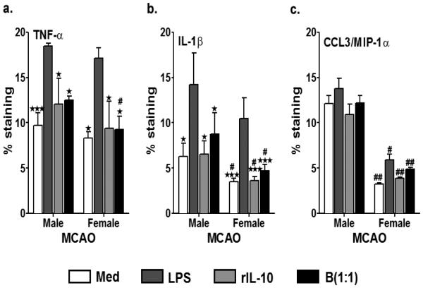
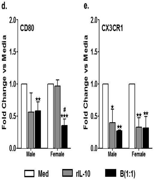
Fig. 4. Enhanced dampening of proinflammatory status of microglia from female mice in response to treatment with IL-10+ B cells, post-MCAO.
Primary MG, isolated and cultured from MCAO-treated WT male and female mice, were harvested after 21 days in vitro (at confluency) and cultured in GM-CSF-free medium for 5 days. MG were stimulated with 10ng/ml LPS for 4 hours. Supernatants were discarded after 4 hours and one of the following treatments was given in 1mL of fresh culture medium: no treatment, 20ng rIL-10 or IL-10+ B cells at a 1:1 ratio with MG. The MG cells were incubated with mentioned treatments at 37°C and 5% CO2 for 24 hours and proinflammatory cytokines a. TNF-α and b. IL-β and c. chemokine CCL3 (MIP-1α) were determined by flow cytometry. Values are given as mean ± S.E.M. Data presented are representative of n = 3, 3 and 2 separate co-culture set-ups, respectively for TNF-α, IL-1β and CCL3 with each treatment condition done in duplicate for every experimental set-up. RNA was isolated from MG from each of the treatments and real time PCR analysis for d. cd80 and e. cx3cr1 were performed. Statistical analysis was performed using One way ANOVA with post-hoc Dunnett test. Significant differences between sample means are indicated as *p≤0.05, **p≤0.01, ***p≤0.001 as compared to the LPS-stimulated condition. Statistical differences between the two sexes were performed by two-way ANOVA followed by the post-hoc Bonferroni multiple comparison test #p≤0.05 and ##p≤0.01, as compared to the respective treatment in male microglia


