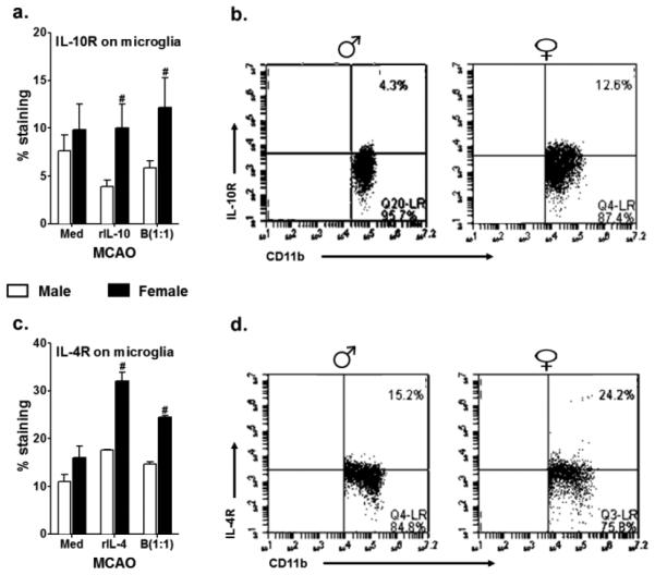
Fig. 6. Increased microglial surface expression of IL-4Rα, and IL-10R by female microglia, post-MCAO.
Primary MG, isolated and cultured from MCAO-treated WT male and female mice, were harvested after 21 days in vitro (at confluency) and cultured in GM-CSF-free medium for 5 days. MG were stimulated with 10ng/ml LPS for 4 hours. Supernatants were discarded after 4 hours and one of the following treatments was given in 1mL of fresh culture medium: no treatment, 20ng recombinant IL-10 (rIL-10), 20ng/ml rIL-4 or IL-10+ B cells at a 1:1 ratio with MG. The MG cells were incubated with mentioned treatments at 37°C and 5% CO2 for 24 hours and A. IL-10 receptor (IL-10R) and C. IL-4Rα were determined by flow cytometry. Representative dot plots indicating increase in the B. IL-10R and D. IL-4Rα in female as compared to male primary MG cultures, post-MCAO. Values are given as mean ± S.E.M. Data presented are representative of n = 2 separate co-culture set-ups, with each treatment condition done in duplicate for every experimental set-up. Statistical differences between the two sexes were performed by two-way ANOVA followed by the post-hoc Bonferroni multiple comparison test, with #p≤0.05 as compared to the respective treatment in male MG

