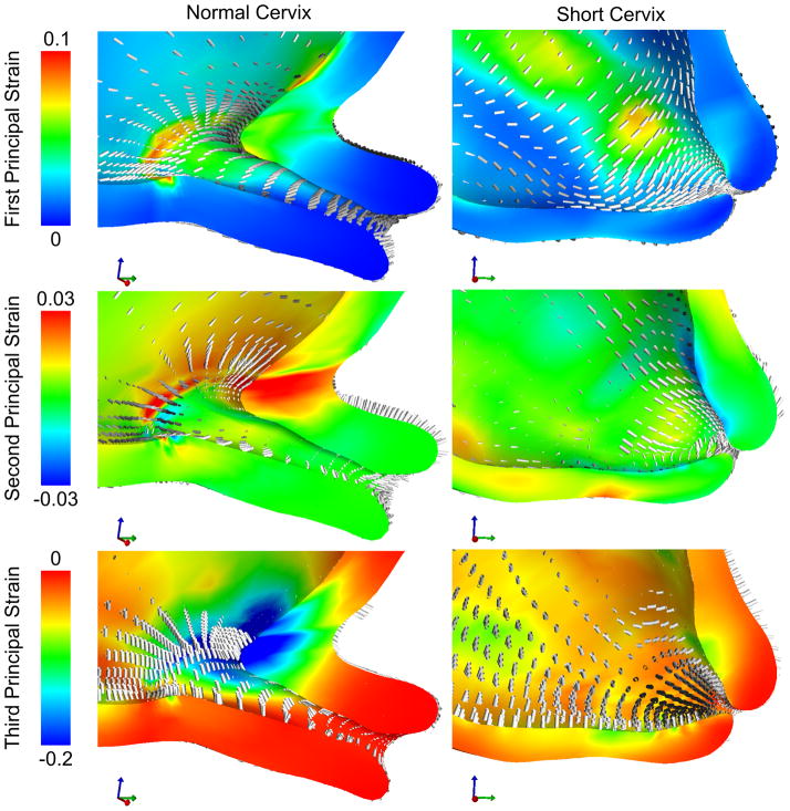
Figure 5.
Strain results for the normal and short cervix model using a random continuous distributed fiber model in the cervix, with an IUP of 8.6 kPa corresponding to the peak IUP during uterine contraction. Circumferential first principal strains around the cervical canal are observed in both models. Strain levels in the short cervix model on the order of 0.04 to 0.06 are present in locations along the whole length of the histological cervical canal. In the normal cervix model, these large strains do not propagate farther than the 1/3 of the cervix closest to the internal os.

