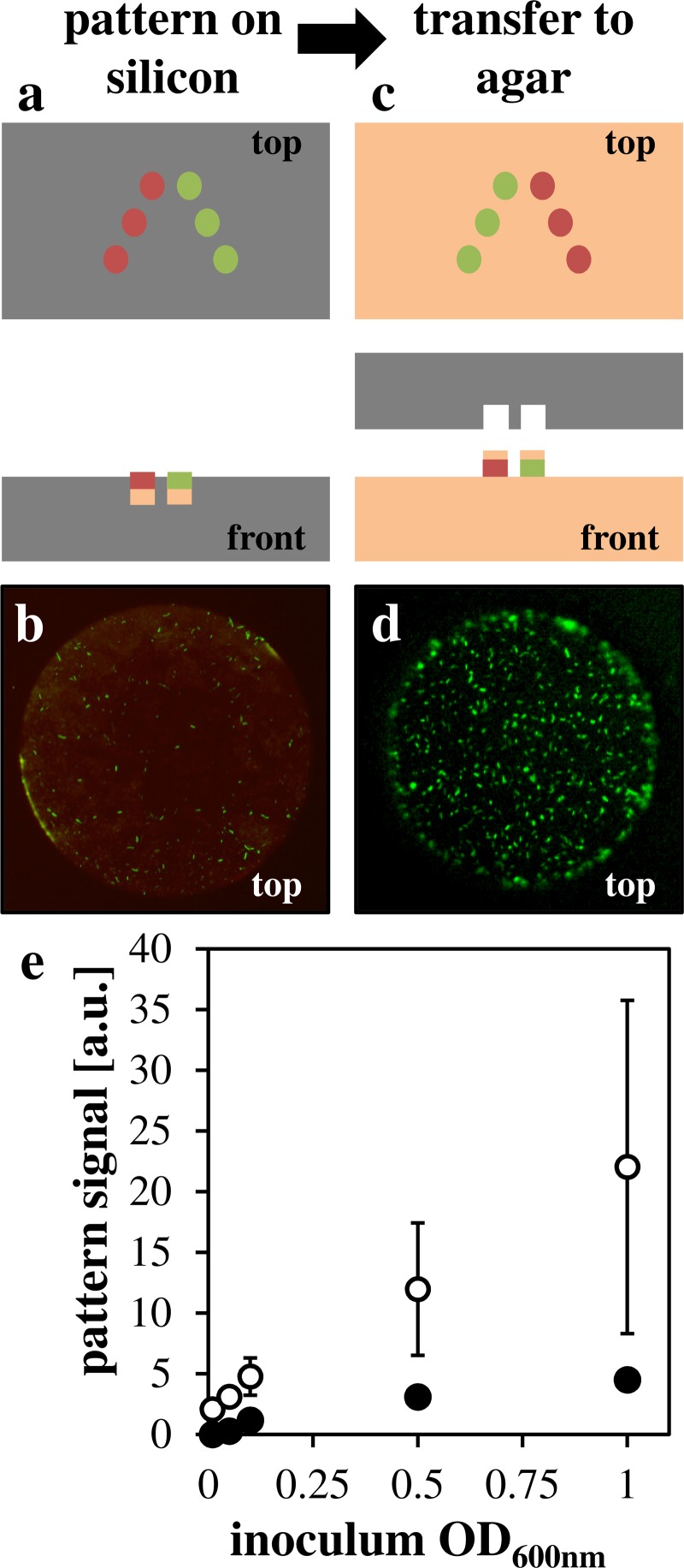
FIG. 3.
Agar pad incorporation for cell transfer. (a) Schematic showing agar pads in silicon microwells (b) representative image of GFP E. coli on RFP-labeled agar pad in silicon well (c) schematic showing agar pad transferred to agar surface (d) representative image of GFP E. coli on agar growth medium with agar pad. (e) Quantification of green pixels on target spot (white points) or off-target background (black points) plotted against OD600 nm.

