Fig. 6.
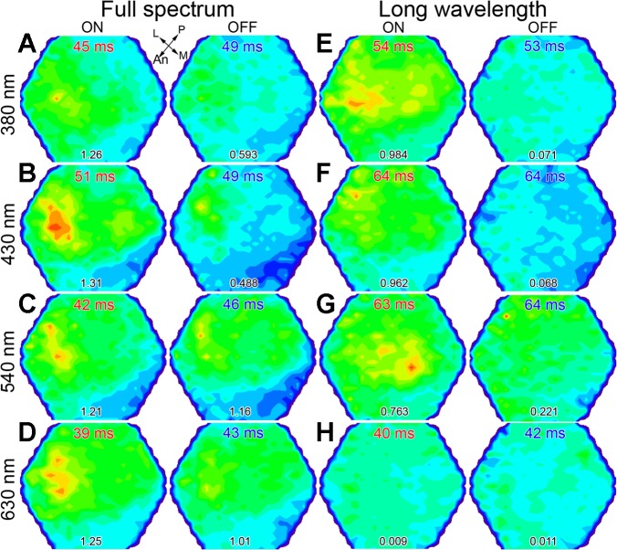
UV cone-mediated sensitivity in the dorsotemporal retina of juvenile rainbow trout. A–H: pairs of pseudocolor images (arranged horizontally) showing maximal ON and OFF responses to different λs (380, 430, 540, or 630 nm) under the dim, full-spectrum light background of Fig. 5 (A–D) and under a >540-nm (long λ) background (E–H), which chromatically adapted the long λ cone mechanism (see minimum signal in H). The numbers at the bottom are the maximal response amplitude in 10−4 ΔF/F. The light guide used to generate the results in Fig. 5, B and C, here, was pointed toward the dorsotemporal retina. Both ON and OFF maxima were located in the anterolateral optic tectum (corresponding to the dorsotemporal retina) under the full-spectrum condition (A–D) and under the long λ background for 380 nm stimulation (E). Under the latter background, the maximal response to 540 nm light shifted toward the medial optic tectum (ventral retina; G). Other presentation of data and abbreviations are as in Fig. 3.

