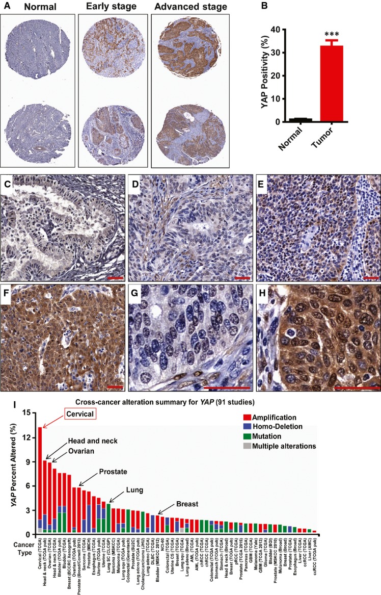
A Representative images show expression of YAP in normal cervical tissues, early-stage cervical cancer (stage I & II), and advanced-stage cervical cancer (stage III & IV). Note the significant increase in YAP immunosignals (brown staining) in the advanced-stage tumors. Tissue core size: 1.5 mm.
B Positivity (percentage of positively stained cells relative to the total number of cells in the tissue section) of YAP immunosignals in the normal and cancerous tissues. Data were analyzed with unpaired t-test in GraphPad Prism 5 with Welch’s correction. Bars represent means ± SEM (n = 10 for normal tissues; n = 69 for tumor tissues; ***P < 0.0001).
C A representative image showing the expression of YAP (brown staining) in normal cervical tissue. Scale bar = 20 μm.
D–F Representative images showing the expression of YAP in (D) early-stage cervical cancer tissue (stage Ib, T1N0M0); (E) medium-stage cervical cancer tissue (stage IIb, T2N0M0); and (F) advanced-stage cervical cancer tissue (stage IIIb, T3N1M0). Scale bar = 20 μm.
G, H High-resolution images showing the cellular location of YAP in (G) early-stage cervical cancer tissue and (H) advanced-stage cervical cancer tissue. Scale bar = 20 μm.
I Multidimensional cancer genomics data analysis results showing cross-cancer YAP gene alteration. The histogram showed the frequencies of YAP gene mutation, deletion, and amplification across cancers. Data were extracted from TCGA database and analyzed using cBioPortal online analyzing tools. The results indicate that the highest alteration frequency of YAP gene occurs in cervical cancer.