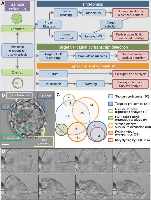
Figure 1. Investigating the blastocoel content using proteomics.

- Study workflow and integration of proteomic, genomic, and embryology experiments.
- The human embryo developed to the blastocyst stage. Five days after fertilization, a human blastocyst shows an inner cell mass (ICM) that will later develop into all the embryonic tissues and a trophectoderm (TE) that will form the extra-embryonic tissues (e.g., the placenta). TE cells are connected and held together by tight junctions that help to contain the fluid within the blastocoel cavity (blastosol). At this stage, the embryo is usually surrounded by a shell of oocyte-derived glycoproteins, the zona pellucida, from which it will “hatch” prior to implantation. Scale bar: 50 μm.
- Embryo usage map. Each circle represents the embryo samples used in specific experimental set (blue, proteomics; orange, embryology; green, gene expression; and red, cytogenetics). Total number per technique is shown in brackets in the legend. In the circles, numbers correspond to the sample used for multiple experiments.
- Sequence of photographs showing progression of the microsuction procedure (blastocentesis). Scale bar: 50 μm.
