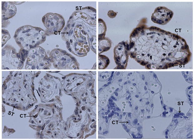
Fig. 4.
Representative results of immunohistochemistry (40×10). The paraffin-embedded placental tissues were sliced into 4 μm sections. The sections were processed as describe under Materials and methods. As negative control (bottom right panel), a section of first-trimester normal placenta was processed with the same procedures except for the absence of primary antibodies. GATAD1 protein was stained brown color. The nuclei were stained blue with haematoxylin. GATAD1 protein localized mostly in the cytoplasm and membrane of syncytiotrophoblasts (ST), and to a less extent, the cytoplasm and membrane of cytotrophoblasts (CT). Higher level of GATAD1 expression was found in third-trimester (upper right) than first-trimester (upper left panel) placenta. Preeclamptic (bottom left) placentas expressed decreased levels of GATAD1 protein compared to normal placentas.

