Fig. 8.
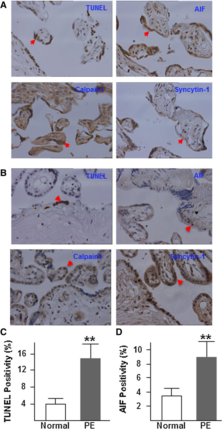
TUNEL and immunohistochemistry analysis of cell apoptosis, and syncytin-1, AIF, and calpain1 in tissue sections from preeclamptic (a) and normal (b) placentas. Brown indicates the staining signals for target factors. The nuclei were counterstained with hematoxylin. Compared to placentas from normal pregnancies, preeclamptic placentas displayed increased cell apoptosis, decreased syncytin-1 expression, increased calpain1, and increased AIF protein expression and nuclear translocation, in syncytiotrophoblasts as well as cytotrophoblasts. Cell counting indicated a significantly higher TUNEL positivity (c) as well as AIF positivity (d) in the preeclamptic placentas (PE) than normal placentas (Normal) (**P < 0.01)

