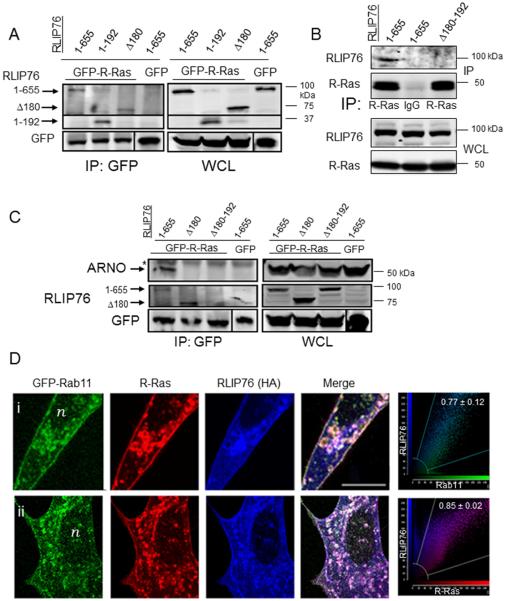
Fig. 1. ARNO and activated R-Ras associate with RLIP76 in a complex at recycling endosomes.
(A) Co-immunoprecipitations (IP) with GFP antibodies from cells expressing GFP-tagged R-Ras(G38V) and HA-RLIP76 truncated or mutant proteins as indicated. (B) Co-IPs of myc-tagged R-Ras(G38V) with HA-tagged RLIP76 variants with α-R-Ras antibodies or control IgG as indicated. (C) Co-IPs of GFP-R-Ras, HA-RLIP76 variants as indicated, and myc-ARNO, with GFP antibodies. *, Ig heavy chain bands in the IPs. (D) Cells from two independent experiments are shown (I, ii). Secondary antibodies used alone as controls showed no detectable staining, and there were no detectable bleedthrough effects across channels (results not shown). Representative two-color component scatter plots comparing the distribution of correlated pixels for Rab11 and RLIP76 (i) or for R-Ras and RLIP76 (ii) and the calculated Pearson's correlation coefficients are shown on the far right. Bar, 10 μm. n, nucleus.

