
Figure 3.
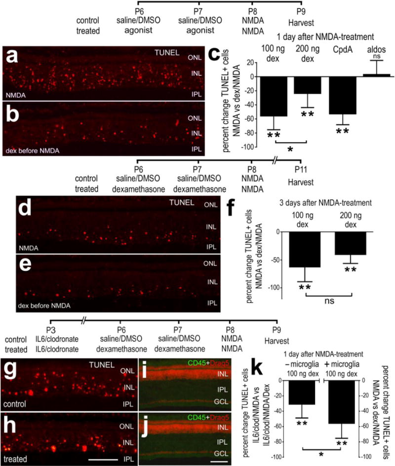
Activation of GCR-signaling before an excitotoxic insult suppresses cell death in NMDA-damaged retinas. Retinas were obtained from eyes that received 2 consecutive daily injections of 100ng/dose Dex (a,b,d,e) or 200ng/dose (c,f), or vehicle at P6 and P7, NMDA at P8, and tissues harvested at P9 or P11. Alternatively, microglia were selectively ablated by using a single intraocular injection of IL6 and clodronate-liposomes at P3 before injections of 100ng Dex/vehicle and NMDA (g–k). Retinal sections were labeled using the TUNEL method (a,b,d,e,g,h), DRAQ5 (red; i,j) and antibodies to CD45 (green; i,j). The scale bar (50 μm) in panel h applies to a,b,d,e,g and h, and the bar in j applies to i and j. c,f,k; The histograms illustrate the mean (±SD; n≥5) percent change in TUNEL-positive cells. Significance of difference (*p<0.05, **p<0.01; ns – not significant) was determined by using Mann-Whitney U test. Abbreviations: ONL – outer nuclear layer, INL – inner nuclear layer, IPL – inner plexiform layer, GCL – ganglion cell layer.