Figure 6.

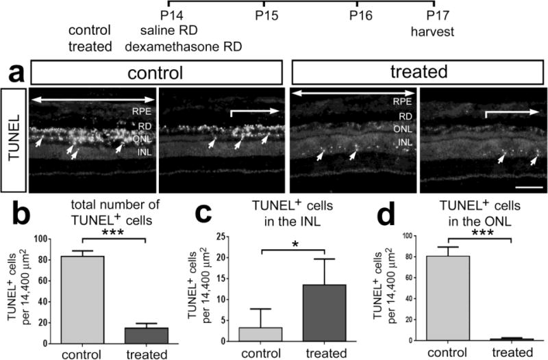
Dex-treatment reduces the loss of photoreceptors following saline-mediated retinal detachment. Retinas were obtained from eyes that received a subretinal injection of saline (control) or saline + soluble Dex (100ng) (treated), and tissues harvested 3 days later. Sections of the retina were labeled using the TUNEL method (a). Large arrows indicate regions of retina where the detachment occurred, and small arrows indicate TUNEL-positive nuclei. Abbreviations: ONL – outer nuclear layer, INL – inner nuclear layer, RPE – retinal pigmented epithelium. Histograms illustrate the mean (±SD) number of TUNEL-positive cells across all layers of the retina (b), INL (c) or ONL (d) in control and Dex-treated retinas. The scale bar denotes 50 μm. Significance of difference (*p<0.05, ***p<0.001, n=4) was determined by using a two-tailed t-test.
