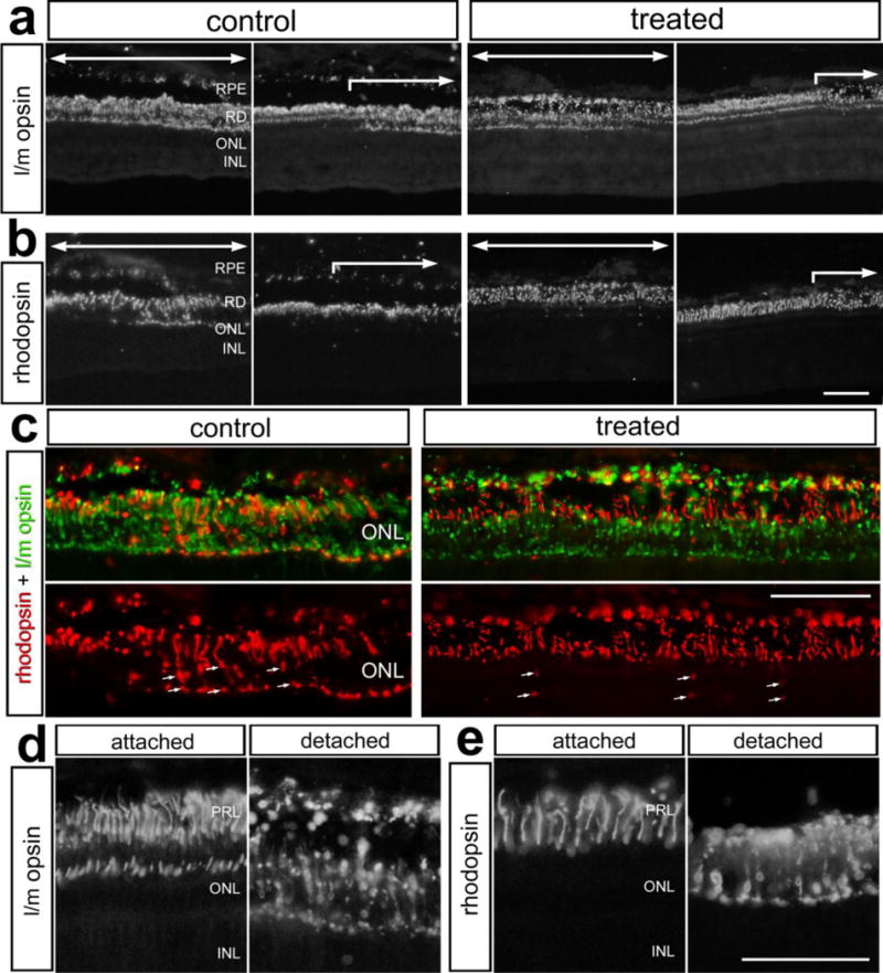
Figure 7.

Dex-treatment reduces the mistrafficking of rhodopsin in rod photoreceptors, but had no effect on mistrafficking of l/m-opsin in cone photoreceptors following retinal detachment. Retinas were obtained from eyes that received a subretinal injection of saline (control) or saline + soluble Dex (treated), and tissues harvested 3 days later. Sections of the retina were labeled using antibodies to l/m-opsin (a,c,d) and rhodopsin (b,c,d). The images in c–e illustrate high-magnification fields of view demonstrating mistrafficking of l/m-opsin and rhodopsin to the inner segments of photoreceptors in control retinas, and in treated retinas in c. Large arrows indicate regions of retina where the detachment occurred. The small arrows indicate mistrafficking of rhodopsin (c). The scale bar (50 μm) in panel b applies to a and b, the scale bar (50 μm) in panel c applies to c, and the scale bar (50 μm) in panel e applies to d and e. Abbreviations: RPE – retinal pigmented epithelium, ONL – outer nuclear layer, INL – inner nuclear layer, IPL- inner plexiform layer, PRL – photoreceptor layer.
