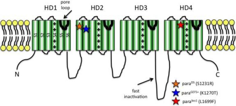
Figure 1. Cartoon depiction of voltage-gated Na+ channel structure.
The channel contains four homology domains (HD1-4) organized around a water-filled pore. Each HD contains six membrane-spanning segments (S1-6). S4 contains positively-charged arginine and lysine residues and acts to sense voltage. An intracellular loop between HD3-4 is responsible for fast inactivation. The S5-6 linker forms the channel pore. The paraGEFS+ and paraDS mutations are located in S1 and S2 of HD2, respectively (orange star, blue star). As described in the text, corresponding mutations in SCN1A cause human epilepsies. The parabss1 mutation is located in S3 of HD4 (red star).

