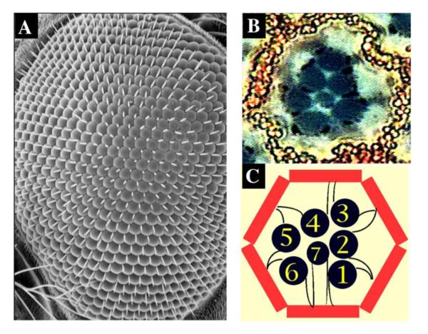
Figure 2. The wildtype Drosophila eye structure.
(A) Scanning electron micrograph of a wild-type adult eye.
(B) Tangential section of one ommatidium unit. High-magnification view. Neuronal photoreceptor cells (black) are surrounded by pigment cells (red).
(C) Illustration of an ommatidium structure. The identity of each photoreceptor cell (black) is labeled. Pigment cells are painted in red.

