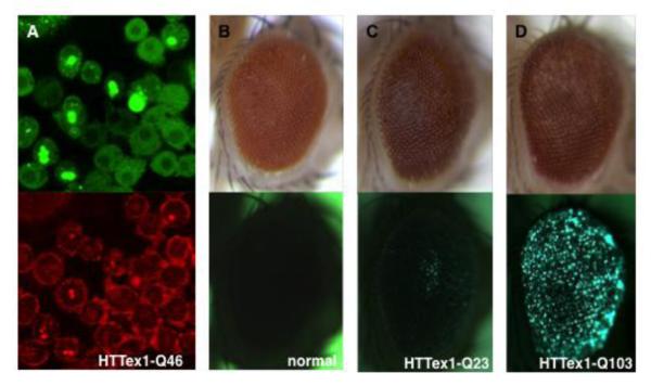
Figure 5. Aggregate formation by mutant HTT in Drosophila.
Formation of aggregates by mutant HTT protein can be modeled and studied in (A) cultured Drosophila cells and (B-D) adult fly eyes. In these studies, mutant HTT exon 1 fragment is revealed by eGFP tag fused in frame at its C-terminus.
(A) A double-labeling image of cultured Drosophila cells that express HTTex1-Q46. Aggregates (bright dots in top picture) are evident in some of the cells. The overall morphology of these cells are marked by staining for cytoskeletal protein F-actin (bottom picture in red), which reveals the sequestration of F-actin in these aggregates (bright dots in bottom picture).
(B-D) Images of same adult fly eyes illuminated by (top panels) bright light to show the overall eye morphology and by (bottom panels) fluorescent light to reveal the presence of eGFP-label HTTex1 aggregates, respectively.
(B) No fluorescent signal in the eye of a wildtype control fly (normal) that did not express human HTT protein.
(C) No clear aggregates in the eye of a transgenic fly that expressed HTTex1 with 23 glutamine (HTTex1-Q23).
(D) Numerous aggregates (bright dots) in the eye of a transgenic fly that expressed mutant HTTex1 with 103 glutamine (HTTex1-Q103).

