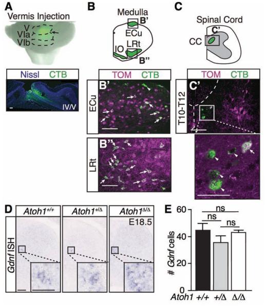
Figure 2. Clarke’s column does not develop predominantly from Atoh1-lineage neurons.
(A-C’) Injection of the retrograde tracer, cholera toxin subunit B (CTB) conjugated to AlexaFluor 488 (green), into the medial area of the adult cerebellum (A, Vermis, Folia V, VIa, VIb, arrow, Nissl Neurotrace 640 in blue) results in backlabeling of cerebellar-projecting neurons in the ECu, LRt, IO (B-B’’), and CC (C-C’). Several Atoh1-lineage neurons (TOM) in the ECu (B’) and LRt (B’’) colabel (arrows) with CTB. However, only 10.5 ± 0.08% of CTB+ cells in CC (C’, arrowheads) colocalize with Atoh1-lineage neurons (arrow)(counts from 18 representative sections from T6-L3 spinal cords per n=2 mice from 2 litters). (D) Gdnf mRNA is unchanged in the Atoh1 wild type (Atoh1+/+, # Gdnf cells, 45 ± 5 ) versus heterozygous (Atoh1+/Δ, 36 ± 5) versus null mice (Atoh1Δ/Δ, 43 ± 2) (p~0.2 to 0.8 in pairwise comparisons, counts from 8 representative T5-11 sections per spinal cord for each genotype, n=3 mice from 3 litters). Mean ± SEM shown. Scale bars are 100 μm and 50 μm for insets in C’ and D. Abbr: external cuneate nucleus (ECu), lateral reticular nucleus (LRt), inferior olive (IO), Clarke’s column (CC), not significant (ns).

