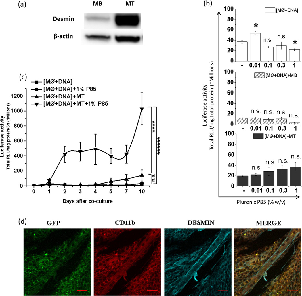
Figure 6. Effect of P85 on horizontal gene transfer from transfected MØs to muscle cells upon co-culture.
(a) Increased desmin expression upon differentiation of MBs to day 7 MTs. (b, c) pDRIVE5Lucia-mDesmin transfected MØs were plated alone, [MØ+DNA], or cocultured with MBs, [MØ+DNA]+MB, or MTs, [MØ+DNA]+MT, and exposed to P85 (0.01, 0.1, 0.3 and 1.0 % (b) or 1 % w/v (c)) for 2 h. Luciferase secreted in cell culture media was quantified after next 24 h (b) or at different time points. In the latter case (c) the media was replaced every 12 h and luciferase secreted in the media over 6 h period was determined daily until day 10. Data represents mean ± SEM, (b) (n=6), (c) (n=12). (b) Statistical comparisons were made using one-way ANOVA with Bonferroni correction for multiple comparisons (c) The AUCs for each individual condition are calculated and compared using one-way ANOVA with Bonferroni correction for multiple comparisons. * p<0.05, n.s. – non significant (d) GFP expression (green) in desmin+ MTs (cyan) was validated 3 days after their coculture with CD11b+ MØs (red) transfected with gWIZ™ GFP pDNA. Transfected MØs were placed on top of MTs. The confocal images were focused at MTs with MØs not visible. The last panels in each row present digitally superimposed images (20 ×) of preceding panels to visualize the co-localization (yellow). Scale bar = 50 µm.

