
Figure 5.
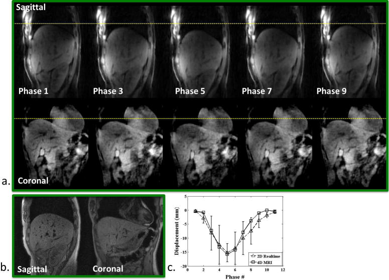
Healthy volunteer. a) Phase-resolved sagittal and coronal images (phase 1, 3, …, 9) reformatted from the 4D MRI image series throughout the entire respiratory cycle. Dashed lines are drawn for better visualization of organ motion at each respiratory phase. b) A single frame of the corresponding real-time 2D-MRI image series. c) Measured SI displacement series, comparing between 4D-MRI and real-time 2D-MRI at each respiratory phase.