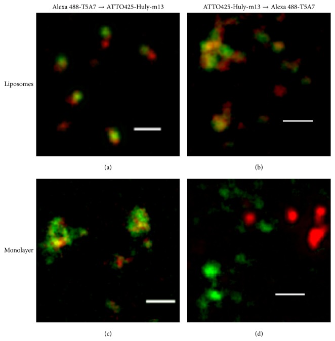
Figure 1.
Stimulated emission depletion (STED) microscopic observation. The LacCer/DOPC liposomes (a, b) and LacCer and DOPC in ethanol (c, d) were coated onto the back surfaces of 96-well NUNC Immunoplates, followed by overnight incubation at room temperature with gentle shaking. The chemical condition of the backside surface of the plate was the same as the surfaces of the wells. The coated plates were blocked with BSA, sequentially stained with Alexa 488-T5A7 (green) ATTO425-Huly-m13 (red) (a, c) or ATTO425-conjugated Huly-m13 (red)→Alexa 488-conjugated T5A7 (green) (b, d), and viewed under a TCS STED CW superresolution microscope (Leica), with signals detected using a GaAsP hybrid detection system (Leica). Deconvolution was performed using Huygens STED deconvolution software (Leica). The panels on the right show enlargements of those on the left. White bars depict 1 μm.

