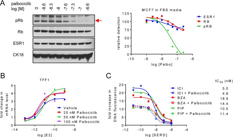
Figure 3. SERDs or SERM/SERD hybrids and CDK4/6 inhibitors impact breast cancer growth by distinct mechanisms.
A) MCF7 cells were plated in media supplemented with FBS prior to 24 hours treatment with increasing concentrations of palbociclib (10−8.6 – 10−6.6 M). Levels of pRb, Rb, ESR1, and CK18 were detected by immunoblot of whole cell extracts (left) followed by densitometry analysis and normalization (right) as in Figure 2C. Red arrow (→) indicates protein band corresponding to pRb. Protein levels were normalized to the control (no palbociclib treatment) present in the first lane. B) MCF7 breast cancer cells were treated for 24 hours with increasing concentrations (10−13 – 10−9 M) E2 in the presence of palbociclib (0, 25, 50 or 100 nM). mRNA expression of TFF1 was analyzed as in Figure 1. C) Proliferation of MCF7 cells was analyzed as in Figure 1 after 8 days treatment with 1nM E2 as well as increasing concentrations of SSH/SERD (ICI, BZA, or PIP) and palbociclib (0 or 25 nM). Data are representative of at least 3 independent experiments.

