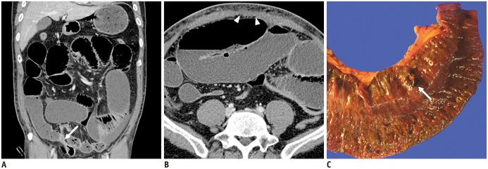
Fig. 6. 47-year-old man presented with abdominal distension for 5 days and dyspnoea for 2 days. He had undergone liposuction 5 days previously at local clinic.
A. On portal venous-phase coronal CT image, abrupt luminal narrowing of small bowel lumen (arrow) is seen. B. Proximal small bowel was diffusely dilated. Focal defect in rectus muscle (arrowheads) was also detected. On diagnostic laparotomy, perfusion was decreased in distal small bowel loop, and segmental resection of ischemic bowel loop was performed. C. Small perforation site was detected, as seen, at resected small bowel loop (arrow). There were also defects in rectus muscle, in sheath below umbilicus, and at liposuction site. Primary repair of these defects was performed during surgery. Despite undergoing emergency operation, patient did not recover from sepsis and died from multi-organ failure.

