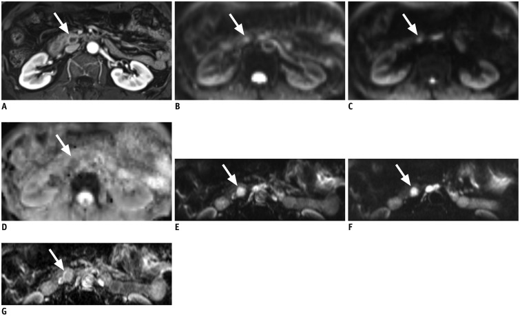
Fig. 1. 58-year-old woman with 1.5 cm sized neuroendocrine tumor (arrow) in pancreas uncinate process.
A. Mass shows rim enhancement on enhanced T1-weighted image. B. Full field-of-view (FOV) diffusion-weighted imaging (DWI) sequence at b = 0 s/mm2. Lesion shows ill-defined hyperintensity. C. Full FOV DWI at b = 500 s/mm2. Note that margin of lesion is severely blurred and barely delineable from background. D. Corresponding apparent diffusion coefficient (ADC) map of full FOV DWI. E. Reduced FOV DWI at b = 0 s/mm2. F. Reduced FOV DWI at b = 400 s/mm2. Lesion is more clearly visualized on reduced FOV image. G. Corresponding ADC map of reduced FOV DWI.

