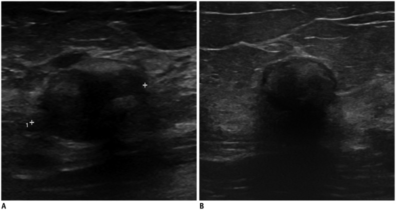
Fig. 2. Fat necrosis in 39-year-old woman without trauma history.
A. Transverse ultrasonography (US) shows irregular hypoechoic mass in her left breast. Lesion measured 2.24 cm in diameter. Pathologically, necrosis was confirmed by US-guided core needle biopsy. B. Follow-up US image after 3 years demonstrates decrease in lesion size to 1.67 cm with increased posterior acoustic shadowing.

