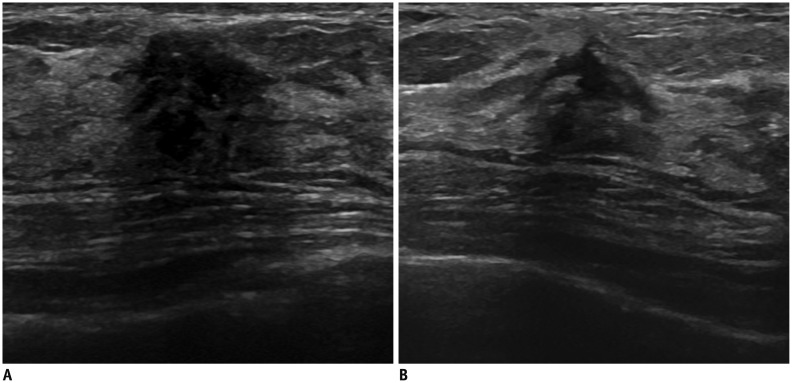
Fig. 3. 43-year-old woman with fibrotic scar.
Initially, she had left breast lesion that was confirmed as fibrocystic change with stromal fibrosis on excisional biopsy (not shown). A. After 5 years of excisional biopsy, lesion shows irregular hypoechoic mass with posterior acoustic shadowing on transverse ultrasonography. Fibrotic nodule with dystrophic calcification was proven from repeated biopsy. B. Transverse ultrasonography image after 7 years shows decreased size of the lesion.

