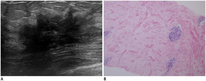
Fig. 6. Diabetic mastopathy in 44-year-old woman with long-standing type 2 diabetes mellitus.
A. Initial transverse ultrasonography image shows irregular spiculated hypoechoic masses with marked posterior shadowing in right breast. B. Photomicrography (hematoxylin and eosin stain, × 100) demonstrates band-like keloid fibrosis with periductal inflammation. After 2 years, lesions remain unchanged on follow-up image (not shown).

