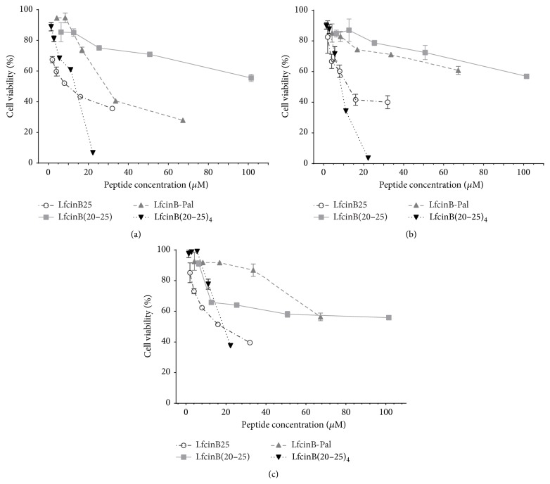
Figure 2.
Cytotoxic effect of LfcinB-derived peptides in the OSCC tumor cell lines CAL27 (a) and SCC15 (b) and the immortalized nontumorigenic keratinocytes cell line Het-1A (c). The cells were incubated for 24 h with the peptides and cell viability was determined by the MTT assay and calculated as the percentage of average absorbance of each treatment relative to the average absorbance of the negative control. The maximum concentration of the peptides used was LfcinB25, 32 μM; LfcinB(20–25), 101.5 μM; LfcinB-Pal, 67.3 μM; LfcinB(20–25)4, 22.25 μM (all equivalent to 100 μg/mL). The data are expressed as the mean ± s.e.m. (n = 3). LfcinB(20–25)4 cf LfcinB(20–25) had statistical significant differences at high concentration (100 μg/mL) (ANOVA, posttest Tukey, p < 0.05).

