
Figure 1.
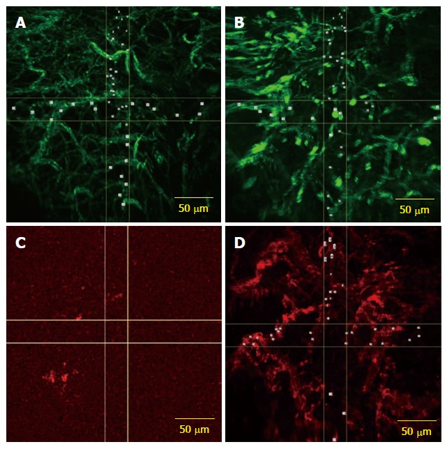
Confocal laser endomicroscopy. A: CLE images with AF488 anti-CD31 antibodies expression on vascular network from both normal; B: Tumor rectal mucosa; C: CLE image showing low expression of the fluorescently labeled anti-CD105 antibodies in normal rectal mucosa; D: Image from the same patient showing microvessels in rectal adenocarcinoma visualized by using CD105 staining as a specific endothelial marker. CLE: Confocal laser endomicroscopy.