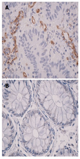
Figure 2.

Immunohistochemistry on CD105 stained sequential sections from rectal cancer tissue samples (magnification 40 ×), CD105-positive vascular endothelial cells were clearly identified by their brown staining (A) and normal rectal mucosa displays the absence of endoglin expression (B).
