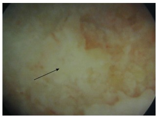
Figure 1.

Endoscopic view of the osteonecrotic lesion. View obtained from the endoscopy through the canal. The posterior aspect of the osteonecrotic lesion is seen at the center of the image (arrow) as a white non-vascularized area, surrounded by normal purple-coloured bone (vascularized bone).
