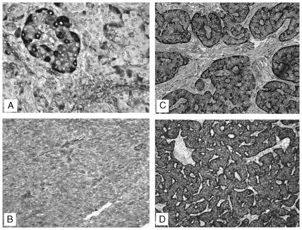
FIGURE 1.
Clusterin expression in nonneoplastic islets and in PNETs. A, Nonneoplastic pancreatic islet: clusterin expression localized mainly at the periphery. B, Neuroendocrine carcinoma: weak intensity with 100% of cells staining. C, Neuroendocrine carcinoma: strong clusterin stain in the basal area of each cell (″secretory″ pattern) and weak stain of the remaining part of the cytoplasm.D, Neuroendocrine carcinoma: strong intensity with 100% of cells staining.

