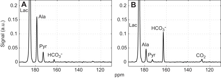
Fig. 1.
Time-averaged 13C signals from rat skeletal muscle after injection of 40 mmol l−1 hyperpolarized [1-13C]lactate. (A) Control. (B) 1 h after DCA infusion. Each spectrum is normalized to the corresponding [1-13C]lactate peak intensity and time averaged for 0–2 min. Relative to the signal intensity of [1-13C]lactate, the DCA stimulation of PDH produces a marked change in 13C spectra, as noted in intensity changes of the alanine, pyruvate, HCO3− and CO2 signals. Scaling all signals to the initial lactate signal intensity corrects for variation in 13C polarization.

