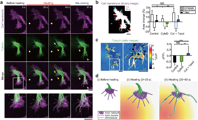
Figure 5. Microtubule elongation and actin-based membrane dynamics in neurites during microheating.
(a) Confocal fluorescence images of CellMask-stained (magenta) neurons expressing tubulin-GFP (green), and merged images. Bottom panels contain magnified versions of the region highlighted in the above merged image. Yellow circles indicate the positions of the heat source. Yellow arrowheads point to areas of microtubule elongation. The T0 was 36 °C, and the laser power was 22 mW. See Supplementary Movie S10. (b) Left: a merged binary image of cell membranes in the absence of chemical agents [Control; same cell as in (a)]. Red binary images (at the first 1 s of heating) and cyan images (after 60 s of heating) are superimposed. Right: relative changes in the surface area of the growth cones in control cells, and in cells treated with 10 μM cytochalasin D (CytoD, see also Supplementary Fig. S12a) or with both 30 μg·mL−1 colchicine (Col) and 20 μM taxol (see also Supplementary Fig. S12b). The relative increases in surface area were compared by one-way ANOVA with Tukey-Kramer tests (*p < 0.05, **p < 0.01; NS, not significant). See also Supplementary Table S3. (c) Left: images depicting the ratio of GFP-tubulin in a control cell after heating to that before heating [same cell as in (a)], and a magnified view of the region highlighted by the white rectangle. Right: relative changes in the fluorescence intensity (ΔF/F0) of GFP-tubulin after heating. The ΔF/F0 values were obtained from the edge of the area of tubulin accumulation within the root of the growth cone before heating (the area surrounded by the grey dashed line in the figures at the left). Due to the microtubule movement from the root toward the tip, there was a decrease in ΔF/F0. See also Supplementary Table S4. The ΔF/F0 values were compared using one-way ANOVA with Tukey-Kramer tests (**p < 0.01, ***p < 0.001; NS, not significant). Scale bars in (b,c), 10 μm. (d) Model of neurite outgrowth during microheating. See text for the detail.

