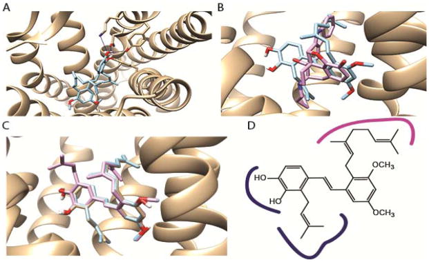
Figure 3.
Docking studies. A. The structure of compound 4 bound in the DOP looking down into the active site from the top. Shows the key proposed H-bonds in blue. B. Analogue 4 (blue) and naltrindole (in pink, from the crystal structure) shown in the active site. C. Lowest energy docking poses of pawhuskin A (1, gray), 3 (pink) and 4 (blue). D. Key features of the pharmacophore of 4 based on the message and address concept of opioid pharmacology, with message region interactions blue and address region interactions pink. The graphics were rendered using the Chimera software suite.

