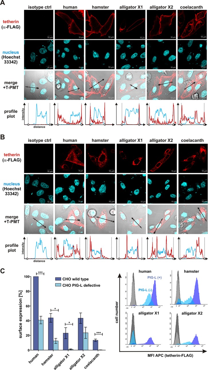
FIG 3.
Subcellular localization and GPI anchor dependency of human, hamster, alligator, and coelacanth tetherin. (A and B) Immunofluorescence pictures of CHO wild-type cells (A) or mutant CHO cells lacking a functional pig-l gene, required for GPI anchor synthesis (B). Two days posttransfection with the indicated tetherin expression vectors, cells were permeabilized and incubated with an anti-FLAG antibody. Nuclei were stained using Hoechst 33342. The regions used to generate profile plots are indicated by black arrows. In the profile plots, the localization of the plasma membrane is indicated by circles and vertical dotted lines. T-PMT, transmission-photomultiplier tube. ctrl, control. (C) Flow cytometric analysis of tetherin levels at the surface of transfected CHO cells (wild type [wt] or PIG-L deficient). Means ± standard errors of the means (SEM) of the results of three to five independent experiments are shown on the left (***, P < 0.001; *, P < 0.05). Examples of primary fluorescence-activated cell sorter (FACS) data indicating the mean fluorescence intensity (MFI) of allophycocyanin (APC) are shown on the right.

