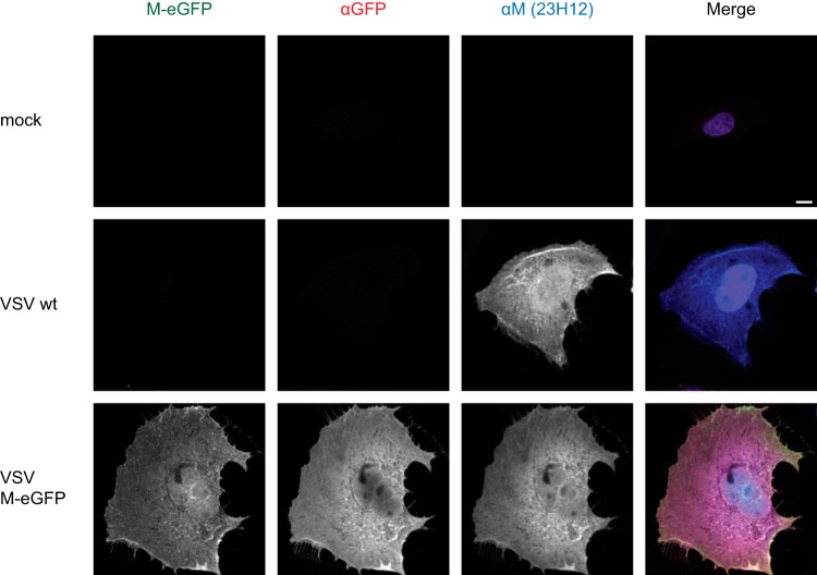
FIG 3.
Subcellular localization of M-eGFP in infected cells. BS-C-1 cells were infected with wild-type VSV or VSV M-eGFP at an MOI of 3, fixed at 8 hpi, and stained with antibodies against eGFP (red) and M (blue). The merged image also includes DAPI (purple). Images were acquired under the same microscope settings and renormalized to the same values; i.e., identical gray value ranges were used for illustration. Bar, 10 μm.

