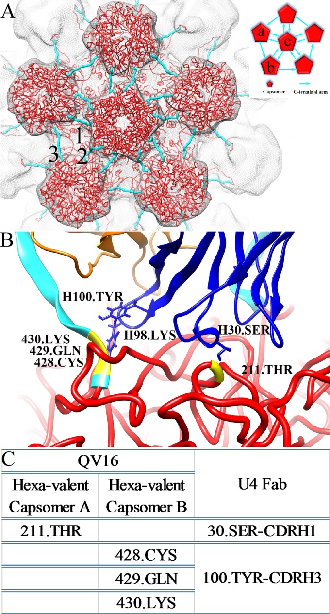
FIG 8.

The H16.U4 epitope. The L1 pseudoatomic structure (red) (PDB ID 3J6R) (18) is fitted into a section of the HPV16 capsid to show the five-fold vertex and the orientations of the C-terminal arms (cyan) that connect capsomers. The inset shows the two hexavalent capsomers (labeled a and b) and one pentavalent capsomer (labeled c) that surround the H16.U4 epitope. Between the capsomers (red pentagons), the direction of the arrows (cyan) indicates the C-terminal arm donor and receiver capsomers as described in the text. (B) The fitted Fab variable domain heavy chains (blue) and light chains (orange) are shown to identify the locations of the contacts between Fab and L1 including positions 428, 429, and 430 (yellow) located on the C-terminal arm (cyan) of the capsomer (capsomer a) and position 211 (yellow) on the shoulder of the capsomer (capsomer b). (C) The residues comprising the epitope are listed in the table, along with predicted binding partners.
