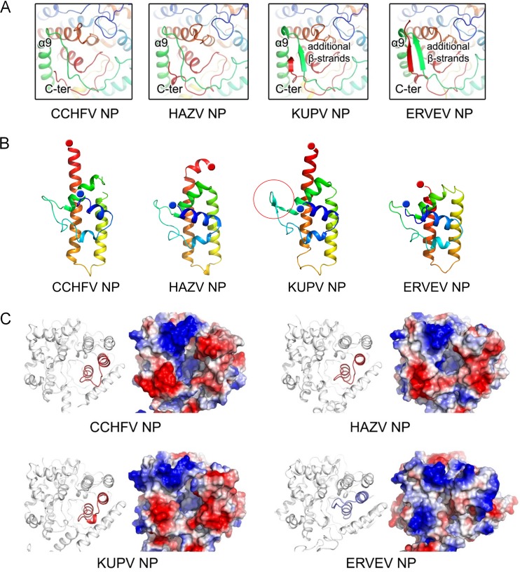
FIG 2.
Structural comparison of nairovirus-encoded NPs. (A) Comparison of the folding of the C terminus and the linkage of α8-α9 in the head domains. All structures are shown as cartoon diagrams in a rainbow color scheme in which the N and C termini are colored blue and red, respectively. (B) Comparison of the stalk domains. A two-β-stranded motif in the KUPV NP stalk domain is highlighted by a red circle. (C) Comparison of the charged pocket of the head domains. The head domain of each NP is shown in a cartoon diagram and a representation of the surface electrostatic potentials.

