
FIG 7.
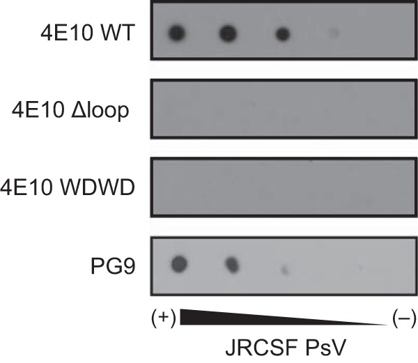
JRCSF PsV recognition as determined by dot blot analysis. Decreasing amounts of PsVs (from left to right) were spotted onto Hybond-C nitrocellulose. The nitrocellulose was then blocked with 5% fat-free milk in PBS (blocking buffer) for 1 h and incubated for 1 additional hour with antibodies (0.2 μg/ml) in blocking buffer at room temperature. The membranes were washed three times for 10 min each time with PBS. Spots were revealed using an HRP-conjugated antibody.