
FIG 4.
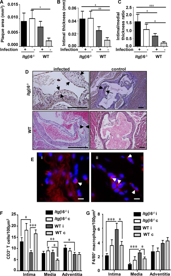
Itgβ6−/− mice exhibit increased aortic plaque accumulation regardless of periodontal infection. (A) Aortic plaque areas. (B) Aortic intimal thicknesses. (C) Aortic intimal layer/medial layer thickness ratios. (D) Representative images of H&E-stained aortic roots from each group. Bar = 50 μm. (E) Representative images of P. gingivalis-positive (red) (white arrowheads) FISH-stained aorta sections within adventitial tissue of infected Itgβ6−/− mice (i) and WT mice (ii). Bar = 10 μm. (F) Aortic CD3+ T cells. (G) Aortic F4/80+ macrophages. All tests were run in triplicates. Data points and error bars represent means ± standard errors of the means for infected (n = 6) and control (n = 6) mice in each group unless stated otherwise (n = 6). i, infected; c, control. *, P < 0.05; **, P < 0.01; ***, P < 0.001 (determined by ANOVA with a Bonferroni posttest). Multiple cross sections were stained and evaluated for measurement and analysis of aortic plaques (2 to 3 sections per aortic area per mouse) by two individuals blind to the treatment groups of the study.