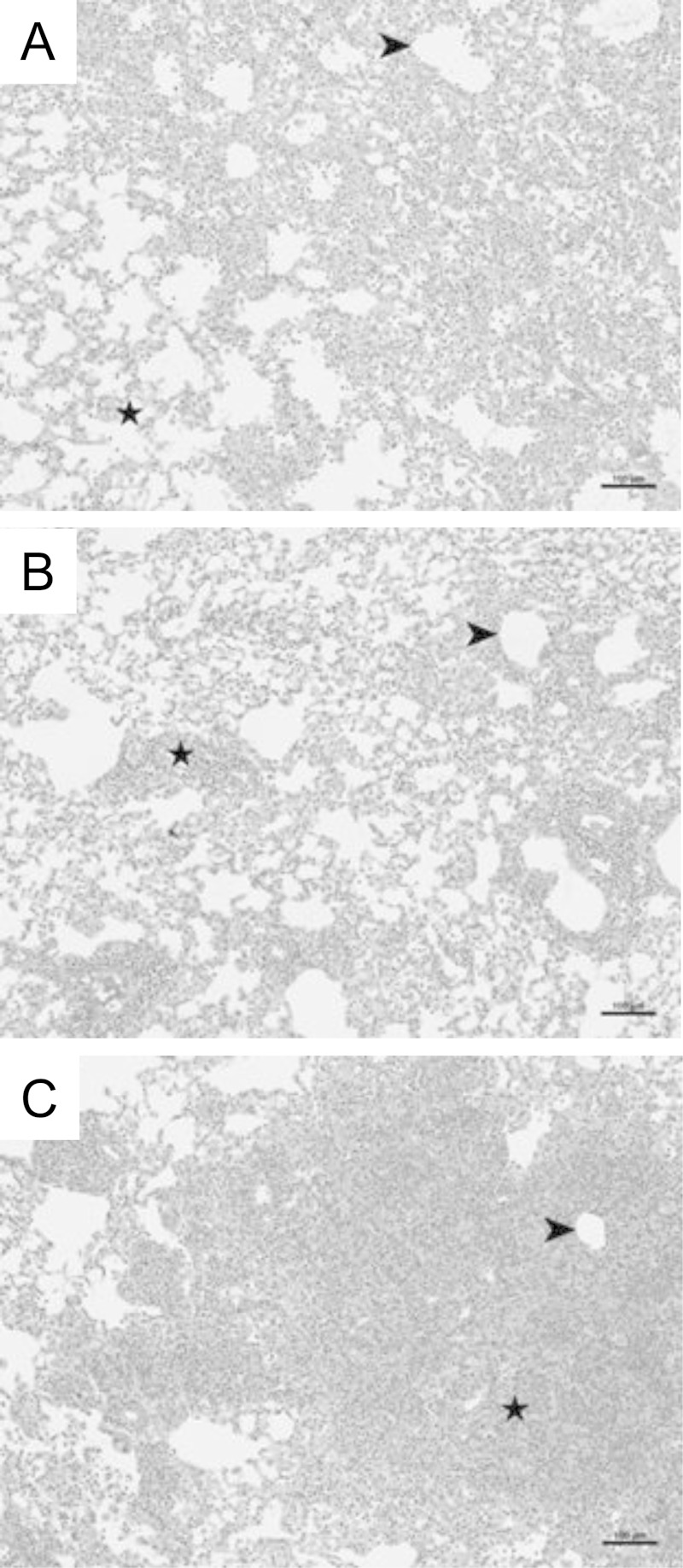
FIG 4.
Loss of IFN-γR signaling is associated with increased lung inflammation. Lung sections were collected from Pneumocystis-infected RAG2−/−, RAG2/IL-4Rα−/−, and RAG2/IFN-γR−/− mice at day 18 post-immune reconstitution and stained with hematoxylin and eosin. Representative pictures were taken at a ×100 magnification. Arrows denote peribronchial regions, and stars denote alveolar regions.

