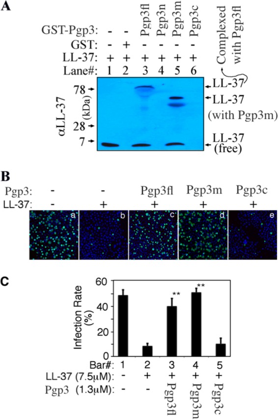
FIG 4.

Mapping the binding domain of Pgp3. (A) Eight micrograms of LL-37 was precipitated alone (lane 1) or with GST agarose beads (lane 2) or GST-Pgp3 full length (Pgp3fl; lane 3), GST-Pgp3 N-terminal region (Pgp3n; lane 4), Pgp3 middle region (Pgp3m; lane 5), or Pgp3 C-terminal region (Pgp3c; lane 6) fusion protein bead in a total volume of 200 μl PBS. After centrifugation, the bead pellets were loaded into the corresponding lanes of an SDS-polyacrylamide gel for Western blot detection with an anti-LL-37 antibody. Note that a portion of LL-37 pulled down by either GST-Pgp3fl or GST-Pgp3m still was complexed with GST-Pgp3fl or GST-Pgp3m even after SDS denaturation. (B) LL-37 at 7.5 μM (equivalent to 30 μg/ml) preincubated without (image b) or with 1.3 μM Pgp3fl (image c), Pgp3m (image d), or Pgp3c (image e) was used to treat C. trachomatis serovar D organisms before the organisms were applied to HeLa cell monolayers. The inclusions were counted as described in the legend to Fig. 1A, and the infection rates were calculated from 3 independent experiments and displayed along the y axis of panel C. Double asterisks indicate statistically significant differences (P < 0.01) between chlamydial cultures treated with LL-37 alone (bar 2) and those treated with LL-37/Pgp3fl or LL-37/Pgp3m complexes (bars 3 and 4).
