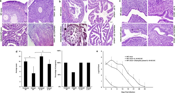
FIG 1.
Histological assessment of genital tract pathology and fertility in Chlamydia-infected mice. (a) Representative hematoxylin and eosin-stained sections of the ovaries of genitally infected WT and IL-10−/− mice and their controls. Changes in tissue morphology were detected in WT mice only. Arrows, examples of vacuolar degeneration of ovarian stromal cells. Bars, 50 μm. (b) Oviduct sections from infected or noninfected WT and IL-10−/− mice. Arrows, regions with vacuolar degeneration of oviductal epithelia. Bars, 50 μm. (c) Representative H&E staining of uteri from infected or noninfected WT and IL-10−/− mice. Arrows, widespread apoptotic necrosis of the endometrium. Bars, 50 μm. (d) Female Chlamydia-infected WT and IL-10−/− mice (n = 10) and their uninfected controls (n = 10) were mated with male mice. The percentage of pregnant mice was determined, and the mean number of pups in the different groups was calculated. Data are presented as means ± SDs. *, P ≤ 0.05 using Student's t test to compare Chlamydia-infected WT and IL-10−/− mice. (e) DCs isolated from IL-10−/− mice were pulsed or not pulsed with live C. muridarum EBs for 2 h and adoptively transferred into female C57BL/6 mice (2.5 × 10⁷ cells per mouse). After 1 week, each mouse was infected intravaginally with 1 × 10⁵ IFU of C. muridarum. The status of the infection was monitored by periodic cervicovaginal swabbing of individual animals and isolation of Chlamydiae in tissue culture. Experiments were repeated twice to give 10 to 12 mice per group. KO, knockout.

