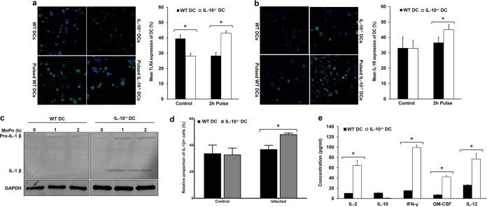
FIG 2.
IL-10 upregulates both innate and adaptive immunity during Chlamydia infection in vitro. (a and b) (Left) Immunofluorescence showing the expression of TLR4 (a) and IL-1R (b) in Chlamydia-pulsed and nonpulsed WT and IL-10−/− DCs. (Right) The graphs shows the percentage of TLR4- and IL-1R-positive DCs ± SD. *, significant difference (P ≤ 0.05) between WT and IL-10−/− DCs. The results are representative of those from three independent experiments. (c) Representative Western blot of pro-IL-1β and active IL-1β expression in supernatants collected from WT and IL-10−/− DCs pulsed with Chlamydia for 0, 1, and 2 h. Note that all samples were run on the same gel and that the gel figure was spliced for better presentation. MoPn, Chlamydia muridarum. (d) Graph showing the percentage of IL-1β-positive Chlamydia-pulsed and nonpulsed WT and IL-10−/− DCs. *, significant difference (P ≤ 0.05) between WT and IL-10−/− DCs. The results are representative of those from three independent experiments. (e) The concentrations of IL-2, IL-10, IL-12, IFN-γ, and GM-CSF secreted from CD4+ T cells cocultured with Chlamydia-pulsed WT or IL-10−/− DCs were measured by a Luminex assay. Data are presented as means ± SDs. *, significant difference (P ≤ 0.05) in the concentration of cytokines from WT and IL-10−/− DCs. The results are representative of those from three independent experiments.

