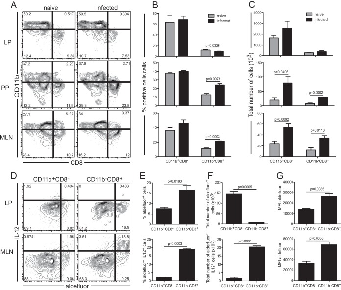
FIG 3.
Responses of mucosal dendritic cell subsets. (A) IL-12p40 YFP mice were orally infected with 2 × 10⁷ E. cuniculi spores, and phenotypic analysis of CD11b+ CD8− and CD11b− CD8+ subsets from the LP, PP, and MLN was conducted at day 2 p.i. Dot plots are gated on MHC-II+ CD11c+ DC. (B and C) Frequency (B) and total number (C) of cells for each DC subset. (D) RALDH activity and IL-12 expression in the CD11b+ CD8− and CD11b− CD8+ subsets from the LP and MLN. Dot plots are gated on the CD11b+ CD8− or CD11b− CD8+ subset from E. cuniculi-infected IL-12p40 YFP mice at day 2 p.i. (E and F) Average frequency (E) or total number (F) of IL-12+ Aldefluor-positive (MLN) or Aldefluor-positive (LP) cells for both the CD11b+ CD8− and CD11b− CD8+ DC subsets (n, 3 to 4 mice/group). (G) MFI of Aldefluor fluorescence for both DC subsets. Data represent results of at least 2 experiments with 3 mice/group.

